
Cellevat3d® nanofiber microcarriers in AAV production

Product specification overview
| Approximate surface area (dry) | 53 000 cm²/g |
| Material | Cellulose nanofiber |
| Surface charge | Cationic |
| Average nanofiber diameter | 500nm |
| Animal free | Yes |
| Typical cell density (HEK293) | 2-3×10⁶ cells/mL |
| Bioreactor | Stirred-tank bioreactors |
| Sterilization procedure | Autoclavation before use |
| Scalability | From lab- to large-scale production |
| Applications | Viral vectors, iPSCs, recombinant proteins, and organoids |
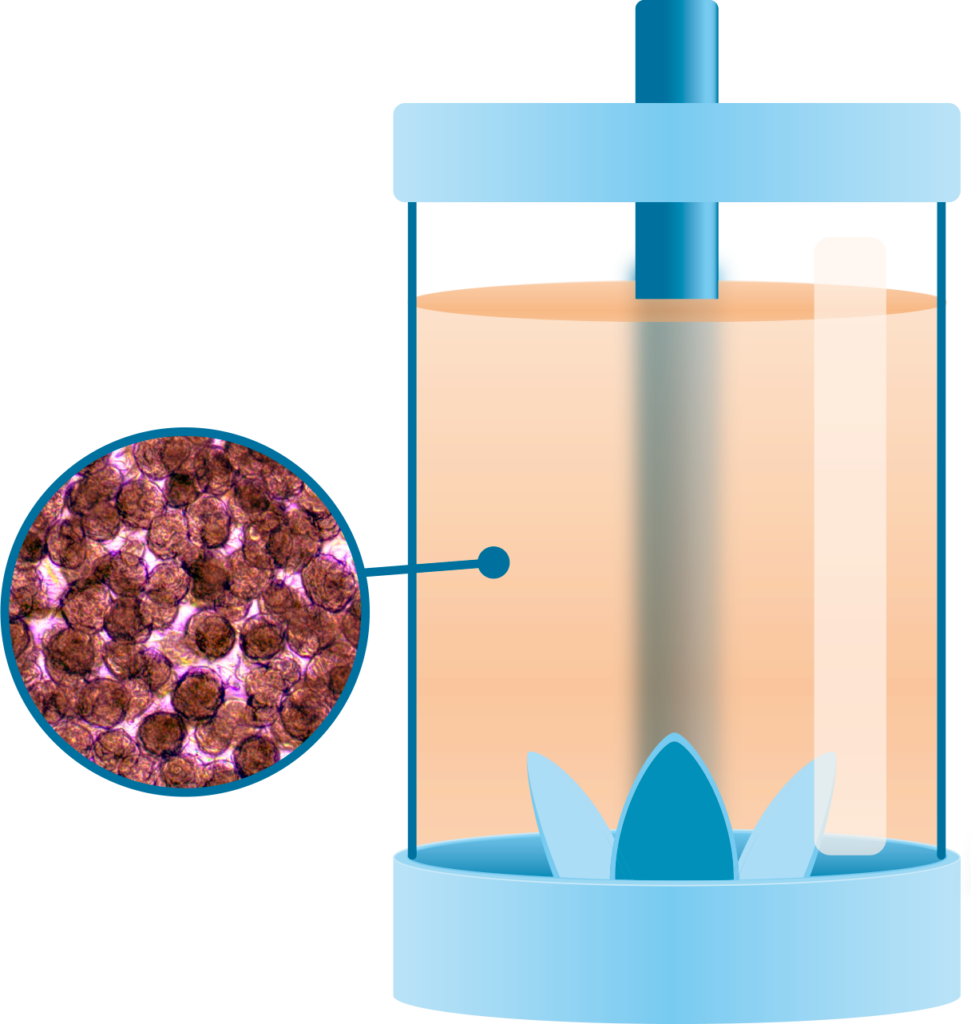
Cellevat3d® nanofiber microcarriers are engineered for stirred-tank bioreactors and are designed using nanofiber cellulose monofibers to support applications such as adeno-associated virus (AAV) and lentivirus (LV) production, induced pluripotent stem cells (iPSCs) production, recombinant protein production, and organoid generation.
Novel nanofiber-based cell culture system for improved upstream productivity in viral vector biomanufacturing
Jessica Giacomoni 1, Anki Knutsson 1, Henrik Bratt 1, Jean-Christophe Bourdon 2, John Counsell 3, Luke Tappouni 3, Mikael Sommarin 4, and Linda Dexlin Mellby 1
1. Cellevate AB, Lund, Sweden, 2. Cellular Medicine, University of Dundee, Dundee, Scotland, 3. UCL Division of Surgery and Interventional Science, London’s Global University, London, UK, 4. Redoxis AB, Lund, Sweden.
Background
Gene therapy and viral vector production are associated with high manufacturing costs that restrict patient access. There is a growing demand for innovative cell culture techniques that improve productivity and scalability. Cellevat3d® nanofiber cell culture systems provide an innovative, sustainable, scalable, and significantly more efficient way for upstream bioprocessing of gene therapy applications.
Results
Data showed that HEK293T cells adhering to Cellevat3d® nanofibers formed homogeneous 3D cultures, generating spheroids with an average diameter of 179 μm after 96h (Fig 1A,B). The scalability of the nanofiber cultures was demonstrated by successful scale-up from spinner flasks (milliliter to liter scale) to a 2.4L Applikon AppliFlex stirred-tank SU bioreactor and a 10L Xcellerex XDR stirred-tank SU bioreactor, maintaining consistent cell growth curves and achieving cell densities up to 3×10⁶ cells/mL, with viability above 95% throughout the culture (Fig 1C,D).
Plasmid transfection showed high transfection efficiency of 88–94% for both AAV2 and AAV9 (Fig 2A,B). AAV2 vectors yielded high volumetric productivity (1014 vg/L) (Fig 3A) with higher titers compared to parallel 2D cultures (Fig 3B). Recent benchmarking results conducted by UCL in London demonstrated a 3.1-fold significant increase in AAV9 vector titer using Cellevat3d® nanofibers compared to current standard microcarriers (Fig 3C).
Cellevat3d® nanofiber-based cell culture systems creates 3D spheroids readily scalable in stirred-tank bioreactors

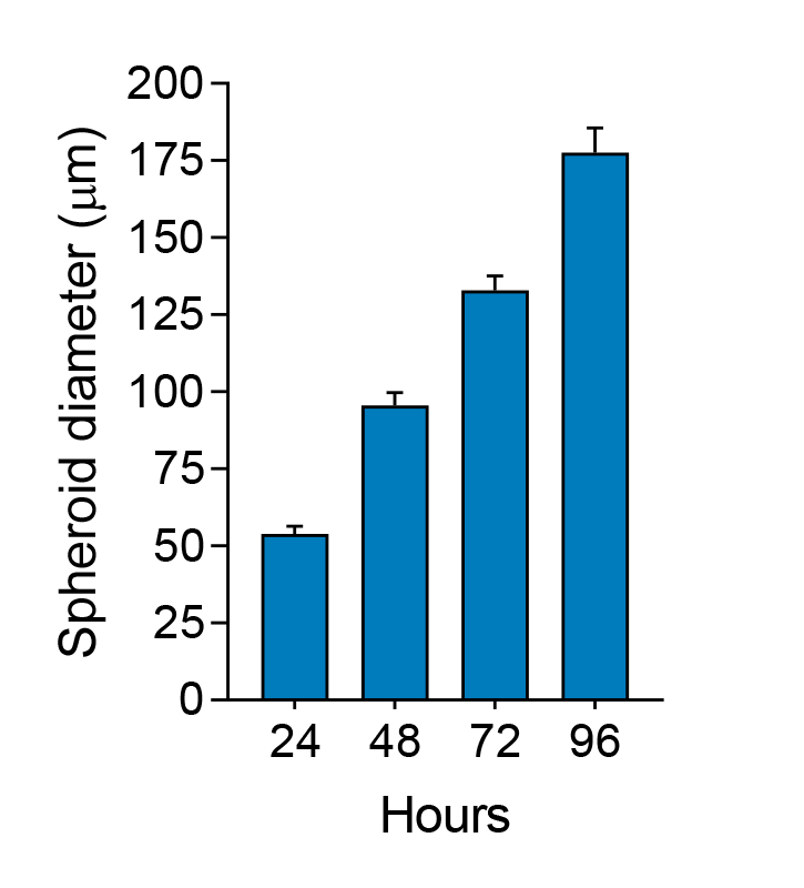

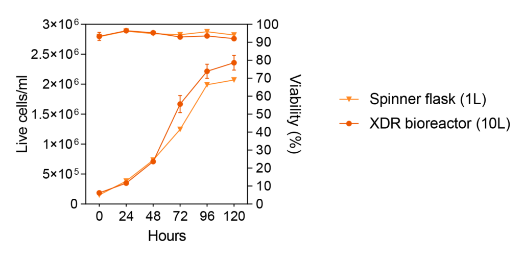
Figure 1. Scalability of Cellevat3d® nanofiber-based cell culture system. A Bright field microscope images of HEK293T cells adhering to nanofibers at inoculum and growing as homogeneous 3D spheroids, shown after 72h in culture. Scale bars 25μm (left top), 50μm (left bottom) and 100μm (right). B Nanofiber cultures (75mL spinner flask) generate 3D spheroids that gradually increase in size over time reaching an average diameter of 179±8 μm after 96h (n=50 measurements per timepoint). C Scale-up from 75mL spinner flask to 1L spinner flask and 2.4L Applikon AppliFlex stirred-tank SU bioreactor resulted in consistent cell growth, reaching yields up to 3×10⁶ cells/mL in 96h. D Scale-up from 1L spinner flask to 10L Xcellerex XDR stirred-tank SU bioreactor maintained similar cell growth trends and an average cell viability of 94% throughout the culture (n=2 independent experiments). Data are presented as either mean or mean ± SEM.
Viral vector production using Cellevat3d® nanofiber cell culture system

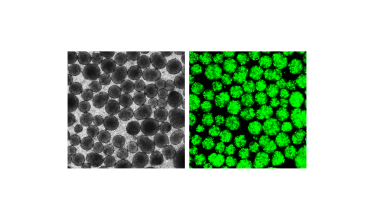
Figure 2. Transient transfection of HEK293T cells on Cellevat3d® nanofibers.
A Plasmid transfection efficiency (%) measured by flow cytometry for AAV2 and AAV9 (n=3-6 biological replicates). B Fluorescence microscope images of ZsGreen-expressing transfected 3D spheroids at 72h post-transfection. Data are presented as mean ± SEM.
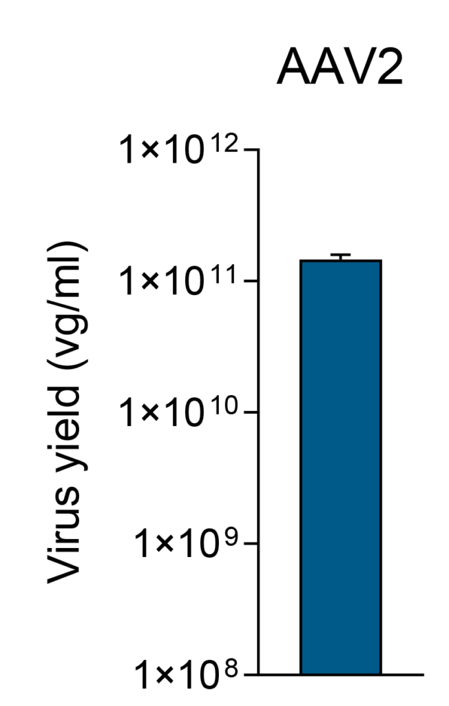
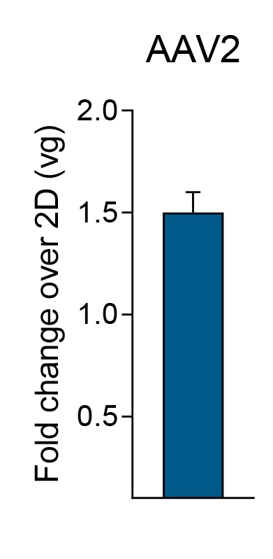
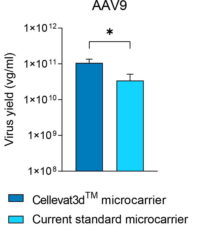
Figure 3. Volumetric productivity of AAV vectors using Cellevat3d® nanofibers. A AAV2 titer of 1.5×10¹¹ vg/mL measured by qPCR (n=4 biological replicates) from 20mL culture.
B Increased product yield (1.5-fold) over parallel AAV2 production using 2D (culture flasks) with same number of cells. C Benchmarking data showed 3.1-fold significant increase in AAV9 titer when using Cellevat3d® nanofibers compared to current standard microcarriers (n=6 biological replicates). Data are presented as mean ± SEM. p* < 0.05, using the Mann–Whitney U test.
Conclusions
The implementation of Cellevate’s new category of nanofiber-based cell culture systems is straightforward and rapid in bioprocess solutions. This product has demonstrated significant benefits for viral vector manufacturing, including successful scale-up to a 10L stirred-tank single-use bioreactor, high transfection efficiency (>90%), and volumetric productivity (1014 vg/L) of human AAV2 vectors. Importantly, benchmarking results revealed a 3.1-fold increase in AAV9 vector titer compared to current standard microcarriers.
These findings highlight Cellevat3d® nanofiber cell culture systems as an innovative, sustainable, scalable, and highly efficient solution for upstream bioprocessing in gene therapy applications.
For more detailed information, download the poster below.
Poster presented at ESGCT, Rome, 22-25 October, 2024