
Cellevat3d® nanofiber microcarriers in lentiviral vector production

Product specification overview
| Approximate surface area (dry) | 53 000 cm²/g |
| Material | Cellulose nanofiber |
| Surface charge | Cationic |
| Average nanofiber diameter | 500nm |
| Animal free | Yes |
| Typical cell density (HEK293) | 2-3×10⁶ cells/mL |
| Bioreactor | Stirred-tank bioreactors |
| Sterilization procedure | Autoclavation before use |
| Scalability | From lab- to large-scale production |
| Applications | Viral vectors, iPSCs, recombinant proteins, and organoids |
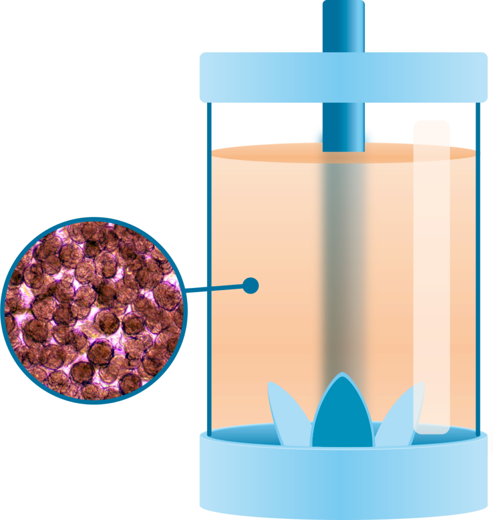
Cellevat3d® nanofiber microcarriers are engineered for stirred-tank bioreactors and are designed using nanofiber cellulose monofibers to support applications such as adeno-associated virus (AAV) and lentivirus (LV) production, induced pluripotent stem cells (iPSCs) production, recombinant protein production, and organoid generation.
Five-fold increase in functional lentiviral vector titers using Cellevat3d® nanofiber microcarriers
Background
Lentiviral vector (LV) manufacturing faces several significant challenges, including high cost of goods (COGs), low to moderate infectious titers and substantial batch-to-batch variability. Many of the current production processes rely on adherent cell cultures, making them labor-intensive, prone to contamination, and not easily scalable.
To address these challenges, Cellevat3d® nanofiber microcarriers offer a cost-effective solution for scaling up adherent cell cultures in controlled stirred-tank bioreactors utilizing an enzyme-free seed-train procedure. This approach reduces batch-to-batch variability, contamination risk, and labor burden. The nanofiber microcarriers, made of sustainable cellulose material, are designed to support the scalable expansion of adherent cells by promoting the formation of homogeneous spheroids, leading to significantly higher virus vector titers and productivity compared to conventional processes.
Results
1. Five-fold increase in functional lentiviral vector titers using Cellevat3d® nanofiber microcarriers compared to industry standard microcarriers
In this study, lentiviral vector production was compared between Cellevat3d® nanofiber microcarriers and industry standard microcarriers. The results demonstrated that nanofiber microcarrier cultures formed homogeneous transfected spheroids with strong EGFP expression (Figure 1A), achieving significantly improved transfection efficiency of 98.6%, compared to 82.4% with industry standard microcarriers (Figure 1B). Importantly, cultures using nanofiber microcarriers produced a 5-fold significant higher infectious titer of 5×10⁷ TU/mL, compared to 1×10⁷ TU/mL with industry standard microcarriers (Figure 1C). To assess production quality, the potency ratio was calculated as infectious titer measured by flow cytometry over physical titer measured by RT-qPCR. Lentiviral vector production using Cellevat3d® nanofiber microcarriers resulted in a markedly higher fraction of infectious particles of 1.7% (Figure 1D).
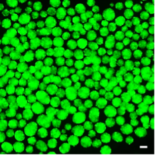
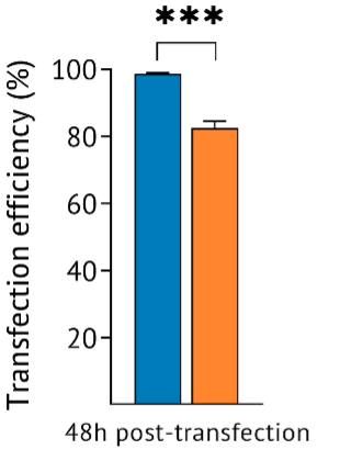
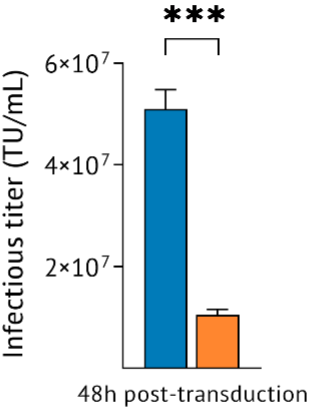
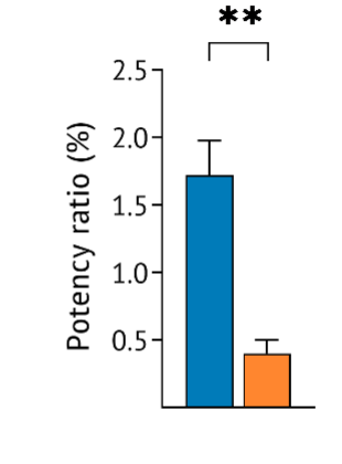

Figure 1. Comparison of lentiviral vector titers using nanofiber and industry standard microcarriers. A Fluorescence microscopy image (4×) of Cellevat3d® nanofiber microcarrier culture expressing EGFP at 48 hours post-transfection. B Improved transfection efficiency of 98.6% vs 82.4% with industry standard microcarrier. Quantification of transfection efficiency (%EGFP-positive cells) at 48 hours post-transfection as analyzed by flow cytometry. p = 0.0003 (unpaired t-test). C 5-fold higher infectious titers vs industry standard microcarriers. Quantification of viral infectious titer at 48 hours post-transduction analyzed by flow cytometry and expressed as transduction units (TU) per mL of culture. p = 0.0002 (unpaired t-test).
D Improved potency ratio of 1.7%. Potency ratio of the crude harvest calculated as the percentage of infectious titer relative to physical titer. p = 0.0094 (unpaired t-test). All data are presented as mean ± SEM from three to four independent experiments.
2. Successful scale-up of Cellevat3d®nanofiber microcarrier cultures into a 10 L bioreactor was achieved using an enzyme-free passaging approach
In a scalability study, enzyme-free passaging was applied by directly transferring Cellevat3d® nanofiber spheroid cultures from 1 L spinner flasks to a 10 L Xcellerex™ XDR single-use (SU) stirred-tank bioreactor (Cytiva). The results showed consistent cell growth curves with an average viability of 94% throughout the culture and reaching cell densities of 2.5×10⁶ cells/mL (Figure 2A).
Furthermore, two sequential cell passages between 125 mL spinner flasks showed consistent cell densities after 96 hours of growth, demonstrating the applicability of the enzyme-free passaging procedure in seed-train processes (Figure 2B).
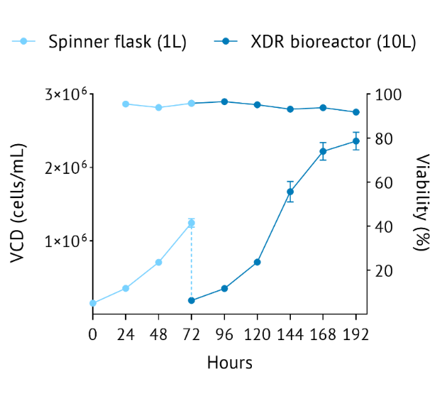
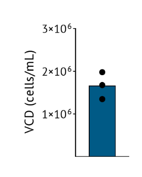
Figure 2. Viable cell density (VCD) and viability of nanofiber microcarrier cultures. A Enzyme-free passaging and scale-up with Cellevat3d® nanofiber microcarriers. HEK 293T spheroid passage from 1 L spinner flask cultured for 72 hours into a 10 L Xcellerex™ XDR SU bioreactor. Data are presented as mean ± SEM from two independent experiments. B VCD of nanofiber microcarrier cultures in 125 mL spinner flask and two sequential cell passages after 96 hours of growth
Conclusions
These findings demonstrate that Cellevat3d® nanofiber microcarriers enable more efficient lentiviral vector production, achieving a 5-fold increase in infectious titer and an improved fraction of infectious particles. Importantly, the enzyme-free passaging procedure provided a scalable and reproducible expansion of HEK 293T cells, streamlining workflows while maintaining high cell quality. In conclusion, Cellevat3d® nanofiber microcarriers offer a sustainable and scalable solution for enhanced lentiviral biomanufacturing productivity.